


18 May 2026
Laboratory-created blood clots: creating a threat to learn how to save lives
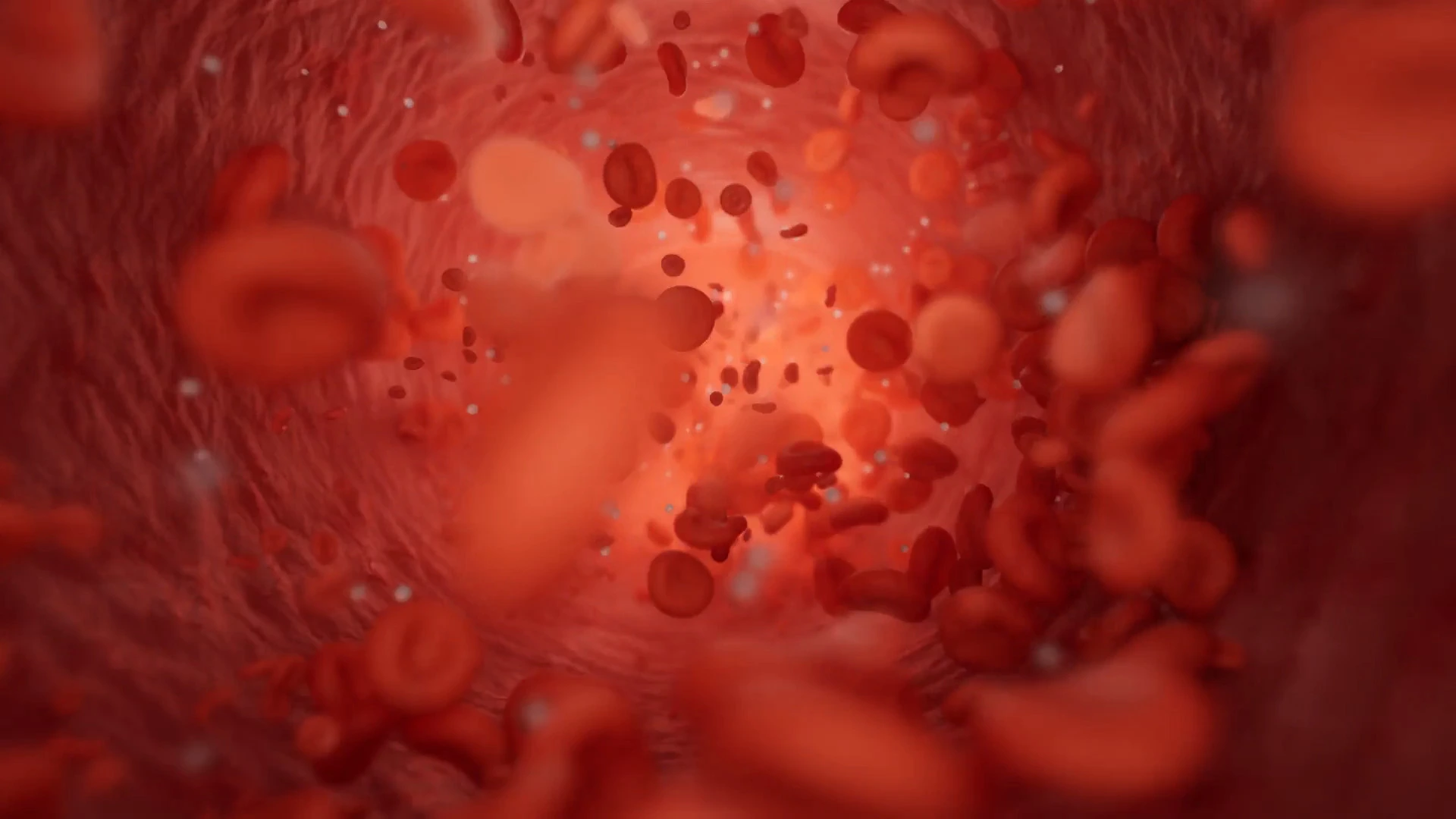
When we hear the word "clot", the first thing that usually comes to mind is not a wound that stops bleeding, but rather its negative side: a stroke, a thrombosis or a heart attack. And that makes sense: blood clots are better known for blocking blood vessels and triggering medical emergencies in a matter of minutes. But that is only a very small part of the story.
A blood clot is also one of the body’s most intelligent responses. When a vessel breaks, the blood needs to react quickly and create a stable structure that protects the tissue while repair begins. In that context, coagulation is not a problem: it is an emergency solution.
That is why a recent study led by McGill University and published in Nature is so interesting. The team has developed a way to create laboratory-designed blood clots, more resistant and adhesive than natural ones, with the aim of controlling severe bleeding and supporting tissue repair. The technique, called click clotting, consists of connecting proteins on the surface of red blood cells through a chemical reaction compatible with the biological environment. According to the authors, these clots are 13 times more resistant to fracture and four times more adhesive than natural ones.
More than a “plug”: how a blood clot is built
To understand why this advance matters, we need to stop imagining a blood clot as a simple "plug" of dried blood. It is a structure that forms, compacts, adapts to the wound and takes part in the beginning of repair.
When an injury occurs, the body acts in stages. First, the blood vessels react and reduce the flow in the damaged area. Then, platelets stick to the wound and create an initial plug. Finally, a protein called fibrin weaves over that plug a kind of mesh that stabilises it and traps blood cells to reinforce it.
For a long time, platelets and fibrin have taken centre stage. But red blood cells also matter: in addition to carrying oxygen, they influence the firmness of the clot and its ability to withstand the pressure of blood flow. And that is where the question raised by this research emerges: if the natural clot can be too fragile in certain situations, can we help the body build a better one?
Click clotting: blood as a biomaterial
McGill’s approach does not involve placing a foreign substance in the body over a wound as if it were just another kind of glue. The proposal is more sophisticated: using the patient’s own red blood cells as structural pieces of a biomaterial.
The technique connects proteins on the surface of those cells through a rapid and biosafe reaction, forming a cellular gel or cytogel in just five seconds. That material then integrates into natural coagulation, becoming embedded within the body’s own fibrin clot.
The key is not only speed, but resistance. In severe bleeding, especially when the area cannot be compressed, the problem is not usually that the body does not know how to clot, but that the clot that forms cannot withstand the pressure of blood flow, detaches or fails to seal the wound properly. A clot that appears quickly, but breaks too soon, may not be enough.
And this is what changes the heart of the matter: it is no longer only about how to stop the flow of blood, but about how to design a structure capable of withstanding its real force inside an organism. The method would allow autologous clots to be prepared using the patient’s own blood in around 20 minutes, or allogeneic clots using compatible donor blood in around 10. A particularly relevant detail if we think about emergency care, surgery or the management of complex wounds.
Clot or thrombus: a matter of context
A natural question when reading about "manufactured clots" is whether this could increase the risk of thrombosis. The answer is simple: the aim is not to introduce clots into the bloodstream, but to create biomaterials that act in a localised context, such as a wound or damaged tissue.
The difference is essential. A pathological thrombus appears where it should not, inside a vessel, and can obstruct flow. A haemostatic clot appears where a lesion needs to be closed. The same biological tool can save or endanger a life depending on the place, the moment and the intensity with which it is activated.
That is why the real advance is not in "manufacturing clots" as a striking concept, but in learning how to regulate their properties: speed, adhesion, resistance, integration with the tissue and ability to support repair. It means moving from observing coagulation to designing it in a more precise and beneficial way.
Blood vessels on a chip: the laboratory imitates the body
Coagulation is difficult to study because it happens quickly, changes with blood flow and depends on many variables. That is why, alongside designed clots, other lines of research are growing, such as blood vessel-on-a-chip models or 3D-printed vessels.
The University of Sydney presented in 2025 a technique to generate anatomical replicas of vessels in just two hours, designed to study stroke-related clots while reducing dependence on animal models. Thromboinflammation-on-a-chip platforms are also being developed, allowing researchers to observe how a clot evolves and resolves in an environment similar to the human body.
All of them point in the same direction: medicine needs more realistic models because the body does not work as a collection of separate parts. Blood, vessels, immune cells, mechanical forces and damaged tissues interact constantly.
From discovery to application
The system has been tested in in vitro trials and rodent models. According to McGill, it improved bleeding control and the regeneration of injured liver tissue, with better performance than the clinical product used for comparison, minimal evidence of immune reactivity and no toxicity in major organs. The researchers themselves stress that more research is still needed before considering clinical use.
The leap from the laboratory to practice is a long one. Researchers will need to study safety in humans, behaviour in different types of wounds, scalability, storage and cost, as well as define which patients would benefit most: people with coagulation disorders, surgeries with a high risk of bleeding, traumatic wounds or liver injuries.
Medicine is always working to find a balance: stopping bleeding without encouraging complications, accelerating repair without causing unwanted responses and reinforcing the natural mechanism without losing control over it.
The precision that healthcare demands
Laboratory-created blood clots remind us that the body does not work in simple categories. What can block flow in a healthy artery can initiate repair in an injured tissue. And that conceptual shift — seeing blood as a biomaterial, giving red blood cells an architectural role and interpreting coagulation as a system that can be precisely reinforced — may be just as important as the technical result.
For areas such as Medicine, Nursing or Bioengineering, this type of advance shows where healthcare is heading: towards more precise solutions, capable of acting at the exact moment, in the right place and with the necessary intensity. At Universidad Mundae, we connect that perspective with a simple idea: studying health is not about memorising isolated processes, but about understanding how they behave when a life is at stake.

